
Page 665 - Atlas of Small Animal CT and MRI
P. 665
Inflammatory Disorders 655
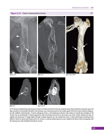
Figure 6.3.2 Chronic Osteomyelitis (Canine) CT
(a) DX, LAT (b) CT, SP (c) CT, 3D, CRAN
(d) CT, TP (e) CT+C, TP
5y FS German Shepherd Dog with previous history of left femoral fracture that was repaired using internal fixation of unknown type. The
dog currently has intermittent left pelvic limb lameness and a draining wound on the medial aspect of the limb, which resolves temporar-
ily with antibiotic administration. A lateral radiograph shows a mid‐diaphyseal malunion with exuberant smooth bony bridging of the
fracture site (a: arrowheads). A central sequestrum with surrounding involucrum is also present (a: arrow). Similar features are seen on
sagittal and transverse CT images (b,d) although multiple sequestra are now evident (d: arrow). A three‐dimensional rendering shows
defects in both the lateral and medial cortices (c: arrows). A contrast‐enhancing tract is seen medial to the femur, which corresponds with
the draining tract later identified clinically (e: arrow).
655