
Page 660 - Atlas of Small Animal CT and MRI
P. 660
650 Atlas of Small Animal CT and MRI
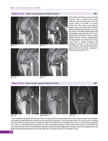
Figure 6.2.14 Cranial Cruciate Ligament Rupture (Canine) MR
5y MC Boxer with history of left pelvic limb
lameness. There is marked joint effusion
within the stifle joint (a,b: open arrow). The
cranial cruciate is not visible in its normal
position (a,c). The caudal cruciate ligament
appears intact with mixed signal intensity,
indicating degenerative change (c,d: arrow).
The menisci are ill defined and irregular with
heterogeneous signal intensity on T1 and T2
images (a,b: arrowhead). A cranial cruciate
ligament rupture, meniscal fragmentation,
and degenerative joint disease were diag-
nosed. There is marked irregularity and
osteophyte formation of the cortical bone
(a) T1, SP (b) T2, SP surrounding the joint. Reproduced with per-
mission from Dr. Silke Hecht, University of
Tennessee, Knoxville, TN, 2014.
(c) T1, SP (d) T2, SP
Figure 6.2.15 Partial Cruciate Ligament Rupture (Canine) MR
(a) T1, SP (b) T2, SPGR, SP (c) ST, DP
11y FS mixed breed with pelvic limb lameness. There is increased soft‐tissue attenuating material within the joint capsule (a: arrowheads),
representing synovial proliferation and effusion. The caudal cruciate ligament is intact (a,b: open arrow). The cranial cruciate is thinned
(a: arrow) and has increased signal on T2 images (b: arrow). The caudal portion of the ligament (not shown) is out of plane but appeared
intact. There is a STIR hyperintense subchondral cyst with surrounding bone edema in the lateral femoral condyle (c: small arrow). This
was distant from the sites of cruciate ligament attachment and was presumed degenerative. A partial cranial cruciate ligament tear was
diagnosed. Reproduced with permission from Dr. Silke Hecht, University of Tennessee, Knoxville, TN, 2014.
650