
Page 658 - Atlas of Small Animal CT and MRI
P. 658
648 Atlas of Small Animal CT and MRI
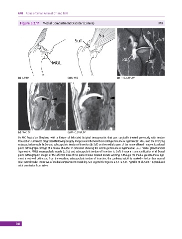
Figure 6.2.11 Medial Compartment Disorder (Canine) MR
(a) IL, MED (b) IL, MED (c) T1+C, ARTH, DP
(d) T1+C, DP (e) T1+C, SPGR, DP
8y MC Australian Shepherd with a history of left‐sided bicipital tenosynovitis that was surgically treated previously with tendon
transection. Lameness progressed following surgery. Images a and b show the medial glenohumeral ligament (a: MGL) and the overlying
subscapularis muscle (b: Su) and subscapularis tendon of insertion (b: SuT) on the medial aspect of the humeral head. Image c is a dorsal
plane arthrographic image of a normal shoulder in extension showing the lateral glenohumeral ligament (c: LGL), medial glenohumeral
ligament (c: MGL), subscapularis muscle (c: Su), and subscapularis tendon of insertion (c: SuT). Image e is a magnification of d. Dorsal
plane arthrographic images of the affected limb of the patient show marked muscle wasting. Although the medial glenohumeral liga-
ment is not well delineated from the overlying subscapularis tendon of insertion, the combined width is markedly thicker than normal
(d,e: arrowheads), indicative of medial compartment instability. See Legend for Figures 6.2.7–6.2.11. Agnello et al 2008. Reproduced
12
with permission from Wiley.
648