
Page 654 - Atlas of Small Animal CT and MRI
P. 654
644 Atlas of Small Animal CT and MRI
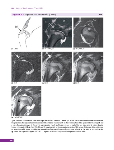
Figure 6.2.7 Supraspinatus Tendinopathy (Canine) MR
(a) IL, MED (b) T1+C, ARTH, SP (c) T1+C, ARTH, TP
(d) ST, SP (e) T2, SP (f) PD, SP
(g) T1+C, ARTH, SP
2y MC Labrador Retriever with acute‐onset right thoracic limb lameness 1 month ago. Pain is elicited on shoulder flexion and extension.
Image a shows the supraspinatus muscle (Ss) and its tendon of insertion (SsT) on the medial surface of the greater tubercle. Images b and
c are arthrographic images of the normal supraspinatus muscle and tendon viewed in sagittal (b) and transverse (c) planes. Sagittal
images of the patient (d–g) show STIR, T2, and PD hyperintensity of the supraspinatus tendon (d–f: arrow). Distension of the joint space
on an arthrographic image highlights the remodeling of the medial aspect of the greater tubercle at the point of tendon insertion
12
(g: arrow). See Legend for Figures 6.2.7–6.2.11. Agnello et al 2008. Reproduced with permission from Wiley.
644