
Page 656 - Atlas of Small Animal CT and MRI
P. 656
646 Atlas of Small Animal CT and MRI
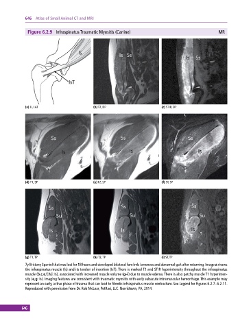
Figure 6.2.9 Infraspinatus Traumatic Myositis (Canine) MR
(a) IL, LAT (b) T2, DP (c) STIR, DP
(d) T1, SP (e) T2, SP (f) ST, SP
(g) T1, TP (h) T2, TP (i) ST, TP
7y Brittany Spaniel that was lost for 18 hours and developed bilateral forelimb lameness and abnormal gait after returning. Image a shows
the infraspinatus muscle (Is) and its tendon of insertion (IsT). There is marked T2 and STIR hyperintensity throughout the infraspinatus
muscle (b,c,e,f,h,i: Is), associated with increased muscle volume (g–i) due to muscle edema. There is also patchy muscle T1 hyperinten-
sity (e,g: Is). Imaging features are consistent with traumatic myositis with early subacute intramuscular hemorrhage. This example may
represent an early, active phase of trauma that can lead to fibrotic infraspinatus muscle contracture. See Legend for Figures 6.2.7–6.2.11.
Reproduced with permission from Dr. Rob McLear, PetRad, LLC. Norristown, PA, 2014.
646