
Page 653 - Atlas of Small Animal CT and MRI
P. 653
Trauma 643
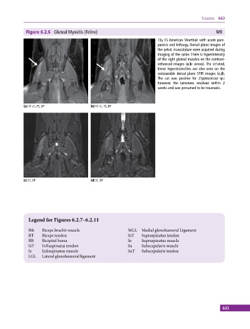
Figure 6.2.6 Gluteal Myositis (Feline) MR
15y FS American Shorthair with acute para-
paresis and lethargy. Dorsal plane images of
the pelvic musculature were acquired during
imaging of the spine. There is hyperintensity
of the right gluteal muscles on the contrast‐
enhanced images (a,b: arrow). The striated,
linear hyperintensities are also seen on the
comparable dorsal plane STIR images (c,d).
The cat was positive for Cryptococcus sp.;
however, the lameness resolved within 2
weeks and was presumed to be traumatic.
(a) T1+C, FS, DP (b) T1+C, FS, DP
(c) ST, DP (d) ST, DP
Legend for Figures 6.2.7–6.2.11
Bib Biceps brachii muscle MGL Medial glenohumeral Ligament
BT Biceps tendon SsT Supraspinatus tendon
BB Bicipital bursa Ss Supraspinatus muscle
IsT Infraspinatus tendon Su Subscapularis muscle
Is Infraspinatus muscle SuT Subscapularis tendon
LGL Lateral glenohumeral ligament
643