
Page 650 - Atlas of Small Animal CT and MRI
P. 650
640 Atlas of Small Animal CT and MRI
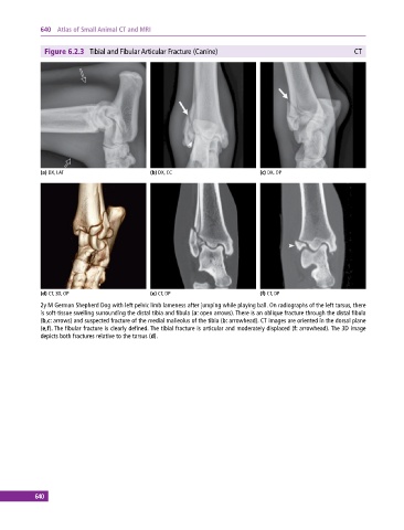
Figure 6.2.3 Tibial and Fibular Articular Fracture (Canine) CT
(a) DX, LAT (b) DX, CC (c) DX, OP
(d) CT, 3D, OP (e) CT, DP (f) CT, DP
2y M German Shepherd Dog with left pelvic limb lameness after jumping while playing ball. On radiographs of the left tarsus, there
is soft‐tissue swelling surrounding the distal tibia and fibula (a: open arrows). There is an oblique fracture through the distal fibula
(b,c: arrows) and suspected fracture of the medial malleolus of the tibia (b: arrowhead). CT images are oriented in the dorsal plane
(e,f). The fibular fracture is clearly defined. The tibial fracture is articular and moderately displaced (f: arrowhead). The 3D image
depicts both fractures relative to the tarsus (d).
640