
Page 655 - Atlas of Small Animal CT and MRI
P. 655
Trauma 645
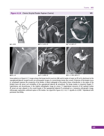
Figure 6.2.8 Chronic Bicipital Tendon Rupture (Canine) MR
(a) IL, MED (b) T1+C, ARTH, SP (c) T1+C, ARTH, TP
(d) DX, LAT (e) T1+C, ARTH, SP (f) T1+C, SPGR, ARTH, TP
Same patient as in Figure 6.2.7. Image a shows the biceps brachii muscle (a: BiB) and its tendon of origin (a: BT) at its attachment to the
supraglenoid tubercle. Images b and c are arthrographic images of a normal biceps tendon (b,c: arrow). Distension of the bicipital bursa
(c: arrowhead) improves the conspicuity of the tendon. A lateral radiograph of the patient shows mineralization in the region of the
bicipital bursa (d: arrow) and remodeling of the supraglenoid tubercle (d: arrowhead). The biceps tendon is irregularly shaped, T1
hyperintense, and discontinuous in the sagittal arthrographic image (e: arrow). T1 hyperintense remnants of the ruptured tendon
(f: arrow) are seen adjacent to the cranial margin of the supraglenoid tubercle (f: arrowhead) on a transverse arthrographic image.
Arthroscopic exploration confirmed rupture of the tendon. See Legend for Figures 6.2.7–6.2.11. Agnello et al 2008. Reproduced with
12
permission from Wiley.
645