
Page 657 - Atlas of Small Animal CT and MRI
P. 657
Trauma 647
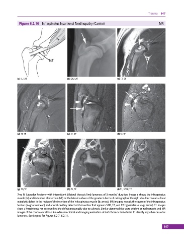
Figure 6.2.10 Infraspinatus Insertional Tendinopathy (Canine) MR
(a) IL, LAT (b) DX, LAT (c) T2, SP
(d) ST, SP (e) ST, DP (f) ST, TP
(g) PD, TP (h) T1, TP (i) T1, SPGR, TP
7mo M Labrador Retriever with intermittent bilateral thoracic limb lameness of 3 months’ duration. Image a shows the infraspinatus
muscle (Is) and its tendon of insertion (IsT) on the lateral surface of the greater tubercle. A radiograph of the right shoulder reveals a focal
osteolytic defect in the region of the insertion of the infraspinatus muscle (b: arrow). MR imaging reveals the course of the infraspinatus
tendon (e–g: arrowhead) and a focal cavitary defect at its insertion that appears STIR, T2, and PD hyperintense (c–g: arrow). T1 images
show a hypointense rim surrounding the defect presumably due to sclerosis. Similar abnormalities were evident on radiographs and MR
images of the contralateral limb. An extensive clinical and imaging evaluation of both thoracic limbs failed to identify any other cause for
lameness. See Legend for Figures 6.2.7–6.2.11.
647