
Page 652 - Atlas of Small Animal CT and MRI
P. 652
642 Atlas of Small Animal CT and MRI
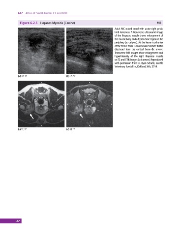
Figure 6.2.5 Iliopsoas Myositis (Canine) MR
Adult MC mixed breed with acute right pelvic
limb lameness. A transverse ultrasound image
of the iliopsoas muscle shows enlargement of
the muscle body and a hypoechoic region in the
periphery (a: calipers). At the lesser trochanter
of the femur, there is an avulsion fracture that is
displaced from the cortical bone (b: arrow).
Transverse MR images show enlargement and
hyperintensity of the right iliopsoas muscle
on T2 and STIR images (c,d: arrow). Reproduced
with permission from Dr. Ryan Schultz, Seattle
Veterinary Specialists, Kirkland, WA, 2014.
(a) US, TP (b) US, SP
(c) T2, TP (d) ST, TP
642