Page 649 - Atlas of Small Animal CT and MRI
P. 649

Trauma  639


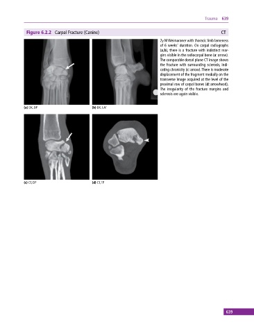
              Figure 6.2.2  Carpal Fracture (Canine)                                                      CT
                                                                             2y M Weimaraner with thoracic limb lameness
                                                                             of 6 weeks’ duration. On carpal radiographs
                                                                             (a,b), there is a fracture with indistinct mar-
                                                                             gins visible in the radiocarpal bone (a: arrow).
                                                                             The comparable dorsal plane CT image shows
                                                                             the fracture with surrounding sclerosis, indi-
                                                                             cating chronicity (c: arrow). There is moderate
                                                                             displacement of the fragment medially on the
                                                                             transverse image acquired at the level of the
                                                                             proximal row of carpal bones (d: arrowhead).
                                                                             The  irregularity  of  the  fracture  margins  and
                                                                             sclerosis are again visible.


            (a) DX, DP                       (b) DX, LAT


            (c) CT, DP                       (d) CT, TP


                                                                                                             639
   644   645   646   647   648   649   650   651   652   653   654