
Page 659 - Atlas of Small Animal CT and MRI
P. 659
Trauma 649
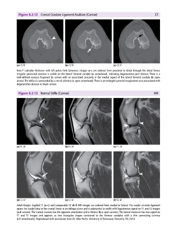
Figure 6.2.12 Cranial Cruciate Ligament Avulsion (Canine) CT
(a) CT, TP (b) CT, TP (c) CT, TP
6mo F Labrador Retriever with left pelvic limb lameness. Images a–c are ordered from proximal to distal through the distal femur.
Irregular periosteal reaction is visible on the lateral femoral condyle (a: arrowhead), indicating degenerative joint disease. There is a
well‐defined osseous fragment (b: arrow) with an associated concavity in the medial aspect of the lateral femoral condyle (b: open
arrow). The defect is surrounded by a rim of sclerosis (c: open arrowhead). There is an enlarged synovial invagination also associated with
degenerative disease (c: black arrow).
Figure 6.2.13 Normal Stifle (Canine) MR
(a) T1, SP (b) T1, SP (c) T1, SP
(d) T2, SP (e) T2, SP (f) T2, SP
Adult Beagle. Sagittal T1 (a–c) and comparable T2 (d–f) MR images are ordered from medial to lateral. The caudal cruciate ligament
spans the caudal tibia to the cranial femur in an oblique plane and is substantial in width with hypointense signal on T1 and T2 images
(a,d: arrows). The cranial cruciate has the opposite orientation and is thinner (b,e: open arrows). The lateral meniscus has low signal on
T1 and T2 images and appears as two triangular shapes contoured to the femoral condyles with a thin connecting isthmus
(c,f: arrowheads). Reproduced with permission from Dr. Silke Hecht, University of Tennessee, Knoxville, TN, 2014.