
Page 675 - Atlas of Small Animal CT and MRI
P. 675
Neoplasia 665
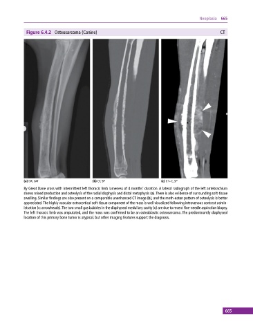
Figure 6.4.2 Osteosarcoma (Canine) CT
(a) DX, LAT (b) CT, SP (c) CT+C, SP
8y Great Dane cross with intermittent left thoracic limb lameness of 4 months’ duration. A lateral radiograph of the left antebrachium
shows mixed production and osteolysis of the radial diaphysis and distal metaphysis (a). There is also evidence of surrounding soft‐tissue
swelling. Similar findings are also present on a comparable unenhanced CT image (b), and the moth‐eaten pattern of osteolysis is better
appreciated. The highly vascular extracortical soft‐tissue component of the mass is well visualized following intravenous contrast admin-
istration (c: arrowheads). The two small gas bubbles in the diaphyseal medullary cavity (c) are due to recent fine‐needle aspiration biopsy.
The left thoracic limb was amputated, and the mass was confirmed to be an osteoblastic osteosarcoma. The predominantly diaphyseal
location of this primary bone tumor is atypical, but other imaging features support the diagnosis.
665