
Page 677 - Atlas of Small Animal CT and MRI
P. 677
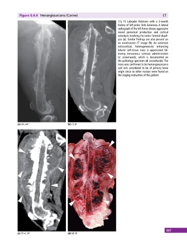
Figure 6.4.4  Hemangiosarcoma (Canine)                                                      CT

                                                                             11y FS Labrador Retriever with a 2‐month
                                                                               history of left pelvic limb lameness. A lateral
                                                                             radiograph of the left femur shows aggressive
                                                                             mixed  periosteal  production  and  cortical
                                                                               osteolysis involving the entire femoral diaph-
                                                                             ysis (a). Similar findings are also present on
                                                                             an  unenhanced  CT  image  (b). An  extensive
                                                                             extracortical,  heterogeneously  enhancing
                                                                             lobular soft‐tissue mass is appreciated fol-
                                                                             lowing intravenous contrast administration
                                                                             (c: arrowheads), which is documented on
                                                                             the pathology specimen (d: arrowheads). The
                                                                             mass was confirmed to be hemangiosarcoma
                                                                             and was considered to be of primary bone
                                                                               origin since no other masses were found on
                                                                             the staging evaluation of the patient.


            (a) DX, LAT                      (b) CT, SP


                                                                                                             667
            (c) CT+C, SP                     (d) GP, SP