
Page 16 - Atlas of Small Animal CT and MRI
P. 16
6 Atlas of Small Animal CT and MRI
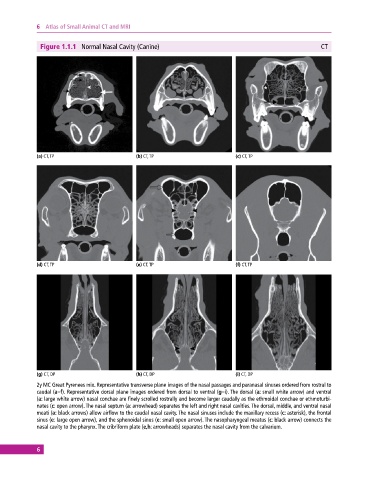
Figure 1.1.1 Normal Nasal Cavity (Canine) CT
(a) CT, TP (b) CT, TP (c) CT, TP
(d) CT, TP (e) CT, TP (f) CT, TP
(g) CT, DP (h) CT, DP (i) CT, DP
2y MC Great Pyrenees mix. Representative transverse plane images of the nasal passages and paranasal sinuses ordered from rostral to
caudal (a–f). Representative dorsal plane images ordered from dorsal to ventral (g–i). The dorsal (a: small white arrow) and ventral
(a: large white arrow) nasal conchae are finely scrolled rostrally and become larger caudally as the ethmoidal conchae or ethmoturbi-
nates (c: open arrow). The nasal septum (a: arrowhead) separates the left and right nasal cavities. The dorsal, middle, and ventral nasal
meati (a: black arrows) allow airflow to the caudal nasal cavity. The nasal sinuses include the maxillary recess (c: asterisk), the frontal
sinus (e: large open arrow), and the sphenoidal sinus (e: small open arrow). The nasopharyngeal meatus (c: black arrow) connects the
nasal cavity to the pharynx. The cribriform plate (e,h: arrowheads) separates the nasal cavity from the calvarium.
6