
Page 19 - Atlas of Small Animal CT and MRI
P. 19
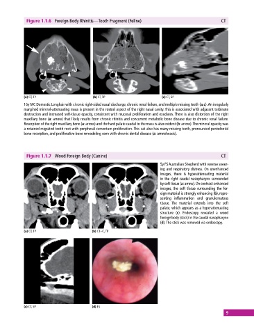
Figure 1.1.6  Foreign Body Rhinitis—Tooth Fragment (Feline)                                 CT


            (a) CT, TP                       (b) CT, TP                       (c) CT, SP
            16y MC Domestic Longhair with chronic right‐sided nasal discharge, chronic renal failure, and multiple missing teeth (a,c). An irregularly
            margined mineral‐attenuating mass is present in the rostral aspect of the right nasal cavity. This is associated with adjacent turbinate
              destruction and increased soft‐tissue opacity, consistent with mucosal proliferation and exudates. There is also distortion of the right
            maxillary bone (a: arrow) that likely results from chronic rhinitis and concurrent metabolic bone disease due to chronic renal failure.
            Resorption of the right maxillary bone (a: arrow) and the hard palate caudal to the mass is also evident (b: arrow). The mineral  opacity was
            a retained migrated tooth root with peripheral cementum proliferation. This cat also has many missing teeth, pronounced periodontal
            bone  resorption, and proliferative bone remodeling seen with chronic dental disease (a: arrowheads).


              Figure 1.1.7  Wood Foreign Body (Canine)                                                    CT

                                                                             5y FS Australian Shepherd with reverse sneez-
                                                                             ing and respiratory distress. On unenhanced
                                                                             images, there  is  hyperattenuating  material
                                                                             in the right caudal nasopharynx surrounded
                                                                             by soft tissue (a: arrow). On contrast‐enhanced
                                                                             images, the soft tissue surrounding the for-
                                                                             eign material is strongly enhancing (b), repre-
                                                                             senting inflammation and granulomatous
                                                                             tissue.  The material extends into the soft
                                                                               palate, which appears as a hyperattenuating
                                                                             structure (c). Endoscopy revealed a wood
                                                                               foreign body (stick) in the caudal nasopharynx
                                                                             (d). The stick was removed via endoscopy.

            (a) CT, TP                       (b) CT+C, TP


            (c) CT, SP                       (d) ES
                                                                                                             9