
Page 22 - Atlas of Small Animal CT and MRI
P. 22
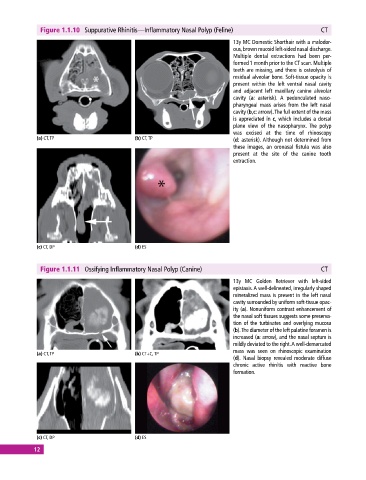
Figure 1.1.10  Suppurative Rhinitis—Inflammatory Nasal Polyp (Feline)                        CT

                                                                              13y MC Domestic Shorthair with a malodor-
                                                                              ous, brown mucoid left‐sided nasal discharge.
                                                                              Multiple dental  extractions  had  been  per-
                                                                              formed 1 month prior to the CT scan. Multiple
                                                                              teeth are missing, and there is osteolysis of
                                                                              residual alveolar bone. Soft‐tissue opacity is
                                                                              present within the left ventral nasal cavity
                                                                              and adjacent left maxillary canine alveolar
                                                                              cavity (a: asterisk).  A pedunculated naso-
                                                                              pharyngeal mass arises from the left nasal
                                                                              cavity (b,c: arrow). The full extent of the mass
                                                                              is appreciated in c, which includes a dorsal
                                                                              plane view of the nasopharynx.  The polyp
                                                                              was excised at the time of rhinoscopy
             (a) CT, TP                       (b) CT, TP                      (d: asterisk). Although not determined from
                                                                              these images, an oronasal fistula was also
                                                                              present at the site of the canine tooth
                                                                              extraction.


             (c) CT, DP                       (d) ES


              Figure 1.1.11  Ossifying Inflammatory Nasal Polyp (Canine)                                   CT
                                                                              13y  MC  Golden Retriever  with  left‐sided
                                                                              epistaxis. A well‐delineated, irregularly shaped
                                                                              mineralized mass is present in the left nasal
                                                                              cavity surrounded by uniform soft‐tissue opac-
                                                                              ity (a). Nonuniform contrast enhancement of
                                                                              the nasal soft tissues suggests some preserva-
                                                                              tion of the turbinates and overlying mucosa
                                                                              (b). The diameter of the left palatine foramen is
                                                                              increased (a: arrow), and the nasal septum is
                                                                              mildly deviated to the right. A well‐demarcated
                                                                              mass was seen on rhinoscopic examination
             (a) CT, TP                       (b) CT+C, TP
                                                                              (d). Nasal biopsy revealed moderate diffuse
                                                                              chronic active rhinitis with reactive bone
                                                                              formation.


             (c) CT, DP                       (d) ES
            12