
Page 25 - Atlas of Small Animal CT and MRI
P. 25
Nasal Cavity and Paranasal Sinuses 15
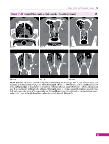
Figure 1.1.15 Mycotic Rhinosinusitis and Osteomyelitis—Aspergillosis (Canine) CT
(a) CT, TP (b) CT, TP (c) CT, TP
(d) CT, DP (e) CT, DP (f) CT, DP
11y MC Rottweiler with chronic left‐sided mucopurulent and hemorrhagic nasal discharge. There is nasal turbinate atrophy with
associated residual mucosal hypertrophy in the left nasal cavity (a,e,f: asterisk). The left frontal sinus contains a soft‐tissue mass with
entrapped fragmented gas (c: large arrow). A mixed pattern of frontal bone osteolysis and periosteal reactive productive response is also
seen (b–e: arrowheads). A focal defect in the left dorsal cribriform plate is seen on both transverse and dorsal plane reformatted images
(c,d: small arrow), and a second defect is suspected on the right side (d: open arrow), although there is no overt nasal disease adjacent
to the cribriform plate on the right. Nasal biopsy confirmed a diagnosis of mycotic rhinosinusitis.
15