
Page 28 - Atlas of Small Animal CT and MRI
P. 28
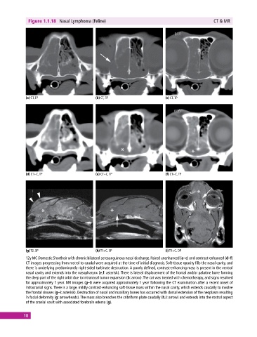
Figure 1.1.18  Nasal Lymphoma (Feline)                                                 CT & MR


             (a) CT, TP                       (b) CT, TP                       (c) CT, TP


             (d) CT+C, TP                     (e) CT+C, TP                     (f) CT+C, TP


             (g) T2, SP                       (h) T1+C, SP                     (i) T1+C, DP
             12y MC Domestic Shorthair with chronic bilateral serosanguinous nasal discharge. Paired unenhanced (a–c) and contrast‐enhanced (d–f)
             CT images progressing from rostral to caudal were acquired at the time of initial diagnosis. Soft‐tissue opacity fills the nasal cavity, and
             there is underlying predominantly right‐sided turbinate destruction. A poorly defined, contrast‐enhancing mass is present in the ventral
             nasal cavity and extends into the nasopharynx (e,f: asterisk). There is lateral displacement of the frontal and/or palatine bone forming
             the deep part of the right orbit due to intranasal tumor expansion (b: arrow). The cat was treated with chemotherapy, and signs resolved
             for approximately 1 year. MR images (g–i) were acquired approximately 1 year following the CT examination after a recent onset of
             intracranial signs. There is a large, mildly contrast‐enhancing soft‐tissue mass within the nasal cavity, which extends caudally to involve
             the frontal sinuses (g–i: asterisk). Destruction of nasal and maxillary bones has occurred with dorsal extension of the neoplasm resulting
             in facial deformity (g: arrowheads). The mass also breaches the cribriform plate caudally (h,i: arrow) and extends into the rostral aspect
             of the cranial vault with associated forebrain edema (g).


            18