Page 30 - Atlas of Small Animal CT and MRI
P. 30

20  Atlas of Small Animal CT and MRI


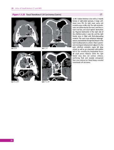
              Figure 1.1.20  Nasal Transitional Cell Carcinoma (Canine)                                    CT

                                                                              7y MC Golden Retriever cross with a 2‐month
                                                                              history of right‐sided epistaxis. A large soft‐
                                                                              tissue mass fills the right nasal cavity and
                                                                              extends across midline (a). The right ectoturbi-
                                                                              nates are obliterated by the mass, and there is
                                                                              right maxillary and nasal septum destruction
                                                                              (a). Regional destruction of the right side of
                                                                              the cribriform plate is seen (b), and the right
                                                                              frontal sinus is filled with fluid‐attenuating
                                                                              material. The nasal mass enhances heteroge-
                                                                              neously and extends through the breach in the
                                                                              right maxillary bone (c: arrow). There is promi-
                                                                              nent meningeal enhancement adjacent to the
                                                                              right  cribriform  osteolytic  region  (d:  large
             (a) CT, TP                       (b) CT, TP
                                                                              arrow) as well as an associated mild midline
                                                                              shift of the interolfactory longitudinal fissure
                                                                              (d: small arrow). Material within the right
                                                                              frontal sinus does not contrast enhance,
                                                                                confirming fluid and exudate entrapment
                                                                              from sinus obstruction. Nasal biopsy revealed
                                                                              transitional cell carcinoma.


             (c) CT+C, TP                     (d) CT+C, TP


            20
   25   26   27   28   29   30   31   32   33   34   35