
Page 35 - Atlas of Small Animal CT and MRI
P. 35
Nasal Cavity and Paranasal Sinuses 25
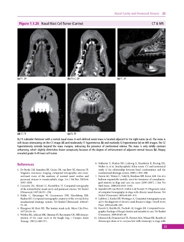
Figure 1.1.26 Nasal Mast Cell Tumor (Canine) CT & MR
(a) T1, DP (b) T1+C, DP (c) T1, SP
(d) CT, TP (e) T2, TP
8y FS Labrador Retriever with a rostral nasal mass. A well‐defined ovoid mass is located adjacent to the right nares (a–c). The mass is
soft‐tissue attenuating on the CT image (d) and moderately T1 hyperintense (b) and markedly T2 hyperintense (e) on MR images. The T2
hyperintensity extends beyond the mass margins, indicating the presence of perilesional edema. The mass is only mildly contrast
enhancing, which slightly diminishes lesion conspicuity because of the degree of enhancement of adjacent normal tissues (b). Biopsy
revealed grade II–III mast cell tumor.
References 6. Schlueter C, Budras KD, Ludewig E, Mayrhofer E, Koenig HE,
Walter A, et al. Brachycephalic feline noses: CT and anatomical
1. De Rycke LM, Saunders JH, Gielen IM, van Bree HJ, Simoens PJ. study of the relationship between head conformation and the
Magnetic resonance imaging, computed tomography, and cross‐ nasolacrimal drainage system. 2009;11:891–900.
sectional views of the anatomy of normal nasal cavities and 7. Berent AC, Weisse C, Todd K, Rondeau MP, Reiter AM. Use of a
paranasal sinuses in mesaticephalic dogs. Am J Vet Res. 2003;64: balloon‐expandable metallic stent for treatment of nasopharyn-
1093–1098. geal stenosis in dogs and cats: six cases (2005‐2007). J Am Vet
2. Losonsky JM, Abbott LC, Kuriashkin IV. Computed tomography Med Assoc. 2008;233:1432–1440.
of the normal feline nasal cavity and paranasal sinuses. Vet Radiol 8. Saunders JH, van Bree H, Gielen I, de Rooster H. Diagnostic value
Ultrasound. 1997;38:251–258. of computed tomography in dogs with chronic nasal disease. Vet
3. Noller C, Henninger W, Gronemeyer DH, Hirschberg RM, Radiol Ultrasound. 2003;44:409–413.
Budras KD. Computed tomography‐anatomy of the normal feline 9. Lefebvre J, Kuehn NF, Wortinger A. Computed tomography as an
nasolacrimal drainage system. Vet Radiol Ultrasound. 2006;47: aid in the diagnosis of chronic nasal disease in dogs. J Small Anim
53–60. Pract. 2005;46:280–285.
4. Hasegawa M, Kern EB. The human nasal cycle. Mayo Clin Proc. 10. Karnik K, Reichle JK, Fischetti AJ, Goggin JM. Computed tomo-
1977;52:28–34. graphic findings of fungal rhinitis and sinusitis in cats. Vet Radiol
5. Webber RL, Jeffcoat MK, Harman JT, Ruttimann UE. MR demon- Ultrasound. 2009;50:65–68.
stration of the nasal cycle in the beagle dog. J Comput Assist 11. Johnson LR, Drazenovich TL, Herrera MA, Wisner ER. Results of
Tomogr. 1987;11:869–871. rhinoscopy alone or in conjunction with sinuscopy in dogs with
25