
Page 39 - Atlas of Small Animal CT and MRI
P. 39
Ear 29
and meningeal contrast enhancement. The scan volume Degenerative disorders
should always include the mandibular and medial
retropharyngeal lymph nodes since reactive lymphade Cartilage mineralization
nopathy and regional metastasis are common. Mineralization of the supportive cartilage of the external
The WHO recognizes squamous cell carcinoma of the ear canal may be seen as an incidental finding but is more
tympanic bulla and tympanic adenocarcinoma as tumors often associated with chronic otitis externa. Mineralization
arising from the middle and inner ear. As with cerumi appears primarily in the horizontal ear canal as linear or
nous adenocarcinomas of the external ear, malignant plaque‐like mineral attenuation on CT (Figure 1.2.18)
tumors of the middle ear may be advanced by the time and may appear as an amorphous signal void in the region
they are imaged, and the specific location of origin may be of the external ear on MRI.
difficult or impossible to determine. Tympanic squamous
cell carcinomas and adenocarcinomas appear similar to Otolithiasis
ceruminous gland carcinomas of the external ear on Otolithiasis of the middle ear has been described in dogs
imaging studies, and differentiation is unlikely. These with active or previous otitis media. Authors ascribed the
tumors are also highly invasive and bone destructive, otoliths to mineralization of necrotic debris in the osse
typically involving the internal ear and often extending ous bulla, but otoliths sometimes appear to arise directly
intracranially (Figures 1.2.16, 1.2.17). Malignant tumors from the internal bulla margins and may well represent
are highly but heterogeneously contrast enhancing on a proliferative osseous response. On CT images, otoliths
both CT and MR images. Pharyngeal and cervical adnexa appear within the tympanic bulla as solitary or multiple
are also frequently affected, and regional lymphadeno mineral densities of variable shape and size (Figure 1.2.19).
pathy is common. Concurrent otitis media may also be seen.
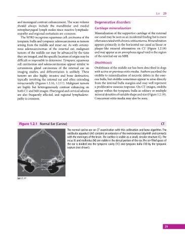
Figure 1.2.1 Normal Ear (Canine) CT
The normal canine ear on CT examination with thin collimation and bone algorithm. The
vestibular aqueduct (AV) contains an extension of the membranous labyrinth and connects
with the meninges of the brain. The cochlea is visible as a small, circular structure (C). The
incus (I) and malleolus (M) are visible in the dorsal portion of the ear. The air‐filled space of
the ear is divided into the tympanic cavity (TC) and tympanic bulla (TB) by the tympanic
septum (not shown).
(a) CT, TP
29