
Page 44 - Atlas of Small Animal CT and MRI
P. 44
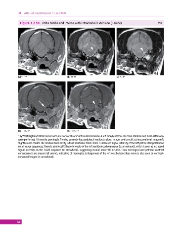
34  Atlas of Small Animal CT and MRI


              Figure 1.2.10  Otitis Media and Interna with Intracranial Extension (Canine)                MR


             (a) T1, TP                       (b) T2, TP                       (c) FL, TP


             (d) T1+C, TP                     (e) T1+C, TP
             13y West Highland White Terrier with a history of chronic otitis externa/media. A left‐sided external ear canal ablation and bulla  osteotomy
             were performed 18 months previously. The dog currently has peripheral vestibular signs. Images a–d are all at the same level. Image e is
             slightly more caudal. The residual bulla cavity is fluid and tissue filled. There is increased signal intensity of the left petrous temporal bone
             on all image sequences. There is also focal T2 hyperintensity of the left vestibulocochlear nerve (b: arrowhead), which is seen as increased
             signal intensity on the FLAIR sequence (c: arrowhead), suggesting cranial nerve VIII neuritis. Focal meningeal and petrosal contrast
             enhancement are present (d: arrow), indicative of meningitis. Enlargement of the left vestibulocochlear nerve is also seen on contrast‐
             enhanced images (e: arrowhead).


            34