
Page 48 - Atlas of Small Animal CT and MRI
P. 48
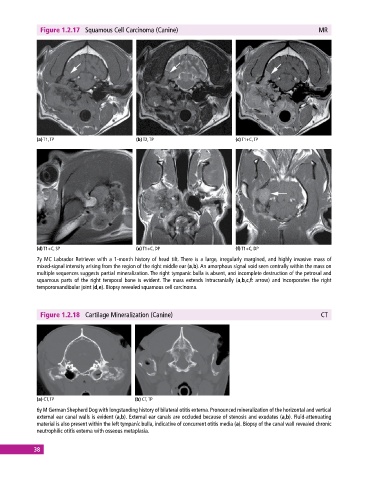
Figure 1.2.17  Squamous Cell Carcinoma (Canine)                                             MR


             (a) T1, TP                       (b) T2, TP                       (c) T1+C, TP


             (d) T1+C, SP                     (e) T1+C, DP                     (f) T1+C, DP
             7y MC Labrador Retriever with a 1‐month history of head tilt. There is a large, irregularly margined, and highly invasive mass of
             mixed‐signal intensity arising from the region of the right middle ear (a,b). An amorphous signal void seen centrally within the mass on
             multiple sequences suggests partial mineralization. The right tympanic bulla is absent, and incomplete destruction of the petrosal and
             squamous parts of the right temporal bone is evident. The mass extends intracranially (a,b,c,f: arrow) and incorporates the right
               temporomandibular joint (d,e). Biopsy revealed squamous cell carcinoma.


              Figure 1.2.18  Cartilage Mineralization (Canine)                                             CT


             (a) CT, TP                       (b) CT, TP
             6y M German Shepherd Dog with longstanding history of bilateral otitis externa. Pronounced mineralization of the horizontal and vertical
             external ear canal walls is evident (a,b). External ear canals are occluded because of stenosis and exudates (a,b). Fluid‐attenuating
               material is also present within the left tympanic bulla, indicative of concurrent otitis media (a). Biopsy of the canal wall revealed chronic
             neutrophilic otitis externa with osseous metaplasia.


            38