
Page 46 - Atlas of Small Animal CT and MRI
P. 46
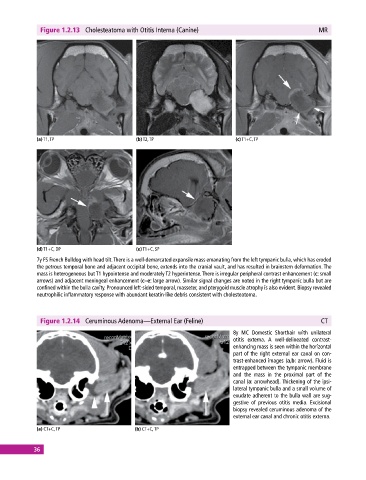
Figure 1.2.13  Cholesteatoma with Otitis Interna (Canine)                                   MR


             (a) T1, TP                       (b) T2, TP                       (c) T1+C, TP


             (d) T1+C, DP                     (e) T1+C, SP
             7y FS French Bulldog with head tilt. There is a well‐demarcated expansile mass emanating from the left tympanic bulla, which has eroded
             the petrous temporal bone and adjacent occipital bone, extends into the cranial vault, and has resulted in brainstem deformation. The
             mass is heterogeneous but T1 hypointense and moderately T2 hyperintense. There is irregular peripheral contrast enhancement (c: small
             arrows) and adjacent meningeal enhancement (c–e: large arrow). Similar signal changes are noted in the right tympanic bulla but are
             confined within the bulla cavity. Pronounced left‐sided temporal, masseter, and pterygoid muscle atrophy is also evident. Biopsy revealed
             neutrophilic inflammatory response with abundant keratin‐like debris consistent with cholesteatoma.


              Figure 1.2.14  Ceruminous Adenoma—External Ear (Feline)                                      CT

                                                                              8y MC Domestic Shorthair with unilateral
                                                                                otitis externa.  A well‐delineated contrast‐
                                                                              enhancing mass is seen within the horizontal
                                                                              part of the right external ear canal on con-
                                                                              trast‐enhanced images (a,b: arrow). Fluid is
                                                                              entrapped between the tympanic membrane
                                                                              and  the mass  in the proximal part  of the
                                                                              canal (a: arrowhead). Thickening of the ipsi-
                                                                              lateral tympanic bulla and a small volume of
                                                                              exudate adherent to the bulla wall are sug-
                                                                              gestive of previous otitis media. Excisional
                                                                              biopsy revealed ceruminous adenoma of the
                                                                              external ear canal and chronic otitis externa.

             (a) CT+C, TP                     (b) CT+C, TP


            36