
Page 45 - Atlas of Small Animal CT and MRI
P. 45
Ear 35
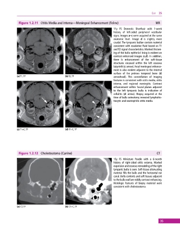
Figure 1.2.11 Otitis Media and Interna—Meningeal Enhancement (Feline) MR
11y FS Domestic Shorthair with 1‐week
history of left‐sided peripheral vestibular
signs. Images a–c were acquired at the same
anatomic level. Image d is slightly more
caudal. The tympanic bullae contain material
consistent with exudative fluid based on T1
and T2 signal characteristics. Marked thicken-
ing of the bulla epithelial lining is evident on
contrast‐enhanced images (c,d). In addition,
there is enhancement of the soft‐tissue
structures encased within the left osseous
labyrinth (c: arrow). Focal meningeal enhance-
ment is also evident adjacent to the internal
surface of the petrous temporal bone (d:
(a) T1, TP (b) T2, TP arrowhead). This constellation of imaging
features is consistent with otitis media, otitis
interna, and regional meningitis. Contrast
enhancement within fascial planes adjacent
to the left tympanic bulla is indicative of
cellulitis (d: arrow). Biopsy acquired at the
time of bulla osteotomy revealed lymphohis-
tiocytic and neutrophilic otitis media.
(c) T1+C, TP (d) T1+C, TP
Figure 1.2.12 Cholesteatoma (Canine) CT
15y FS Miniature Poodle with a 6‐month
history of right‐sided otitis externa. Marked
expansion and osseous remodeling of the right
tympanic bulla is seen. Soft‐tissue attenuating
material fills the bulla and the horizontal ear
canal. Bulla contents and soft tissues adjacent
to the bulla wall are mildly contrast enhancing.
Histologic features of biopsy material were
consistent with cholesteatoma.
(a) CT, TP (b) CT+C, TP
35