
Page 40 - Atlas of Small Animal CT and MRI
P. 40
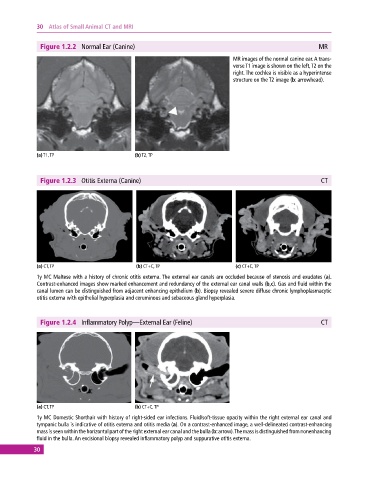
30  Atlas of Small Animal CT and MRI


              Figure 1.2.2  Normal Ear (Canine)                                                           MR
                                                                              MR images of the normal canine ear. A trans-
                                                                              verse T1 image is shown on the left, T2 on the
                                                                              right. The cochlea is visible as a hyperintense
                                                                              structure on the T2 image (b: arrowhead).


             (a) T1, TP                       (b) T2, TP


              Figure 1.2.3  Otitis Externa (Canine)                                                        CT


             (a) CT, TP                       (b) CT+C, TP                     (c) CT+C, TP
             1y MC Maltese with a history of chronic otitis externa. The external ear canals are occluded because of stenosis and exudates (a).
             Contrast‐enhanced images show marked enhancement and redundancy of the external ear canal walls (b,c). Gas and fluid within the
             canal lumen can be distinguished from adjacent enhancing epithelium (b). Biopsy revealed severe diffuse chronic lymphoplasmacytic
             otitis externa with epithelial hyperplasia and ceruminous and sebaceous gland hyperplasia.


              Figure 1.2.4  Inflammatory Polyp—External Ear (Feline)                                       CT


             (a) CT, TP                       (b) CT+C, TP
             1y MC Domestic Shorthair with history of right‐sided ear infections. Fluid/soft‐tissue opacity within the right external ear canal and
             tympanic bulla is indicative of otitis externa and otitis media (a). On a contrast‐enhanced image, a well‐delineated contrast‐enhancing
             mass is seen within the horizontal part of the right external ear canal and the bulla (b: arrow). The mass is distinguished from  nonenhancing
             fluid in the bulla. An excisional biopsy revealed inflammatory polyp and suppurative otitis externa.
            30