
Page 42 - Atlas of Small Animal CT and MRI
P. 42
32 Atlas of Small Animal CT and MRI
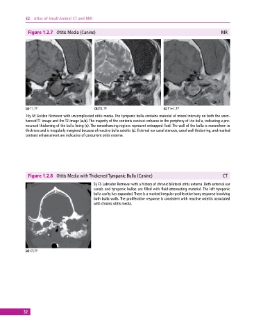
Figure 1.2.7 Otitis Media (Canine) MR
(a) T1, TP (b) T2, TP (c) T1+C, TP
10y M Golden Retriever with uncomplicated otitis media. The tympanic bulla contains material of mixed intensity on both the unen-
hanced T1 image and the T2 image (a,b). The majority of the contents contrast enhance in the periphery of the bulla, indicating a pro-
nounced thickening of the bulla lining (c). The nonenhancing regions represent entrapped fluid. The wall of the bulla is nonuniform in
thickness and is irregularly margined because of reactive bulla osteitis (c). External ear canal stenosis, canal wall thickening, and marked
contrast enhancement are indicative of concurrent otitis externa.
Figure 1.2.8 Otitis Media with Thickened Tympanic Bulla (Canine) CT
5y FS Labrador Retriever with a history of chronic bilateral otitis externa. Both external ear
canals and tympanic bullae are filled with fluid‐attenuating material. The left tympanic
bulla cavity has expanded. There is a marked irregular proliferative bony response involving
both bulla walls. The proliferative response is consistent with reactive osteitis associated
with chronic otitis media.
(a) CT, TP
32