
Page 51 - Atlas of Small Animal CT and MRI
P. 51
Temporomandibular Joint 41
woven bone formation involving the mandible and hyperintense medullary fat with lower intensity tumor
possibly the temporomandibular joints (Figure 1.3.7). (Figures 1.3.14, 1.3.15).
Trauma Degenerative disorders
Injury to the temporomandibular joint is a common Osteoarthrosis
sequela of head trauma. Although conventional radio- Although commonly performed in people because of
graphic imaging can be used to diagnose temporomandib- the high incidence of debilitating degenerative temporo-
ular injury, it consistently underestimates the severity of mandibular joint disorders, there are few reports on the
trauma, particularly when complex fractures are present. use of high‐resolution CT and MR imaging for diagnosis
Luxations and fractures are well delineated using CT imag- of this disorder in veterinary medicine. Although
ing, with imaging features dependent on the specific articular cartilage and the articular disc should be well
trauma sustained (Figures 1.3.8, 1.3.9, 1.3.10, 1.3.11). 9 visualized by MR using appropriate coils and pulse
sequences, MR features of degenerative temporoman-
Inflammatory disorders dibular joint disease have not been fully described in
dogs and cats. CT imaging features include narrowing
Septic arthritis and osteomyelitis of the temporoman- of the joint space (best seen on sagittal plane reformatted
dibular joint are occasionally encountered as a result of images), condyloid process remodeling, subchondral
extension of otitis externa/media or a direct penetrating bone sclerosis, and periarticular new bone formation
injury and may include articular cartilage and subchon- (Figure 1.3.16). Similarly, MR imaging findings may
dral bone destruction, joint distension, and surrounding include joint space narrowing and subchondral bone
cellulitis (Figure 1.3.12). General features of septic and periarticular new bone signal void.
10
arthritis are described in Chapter 6.3.
Ankylosis
Neoplasia
Occasionally, periarticular productive remodeling may
Although uncommon, neoplasia involving the temporo- be exuberant enough to restrict temporomandibular
mandibular joint may arise from intrinsic structures joint range of motion. This can be due either to primary
of the joint or from encroachment from adjacent neo- temporomandibular degenerative joint disease or an
plasms. Benign bone tumors, such as osteomas that arise adjacent proliferative response of the temporal bone
from the mandible or temporal bone, may impinge on associated with chronic otitis. True ankylosis is defined
the temporomandibular joint and will typically appear as bone fusion or synostosis. Most patients with reduced
as a dense, well‐delineated mass on CT and as a low or range of motion, in fact, have extracapsular or fibrous
no signal intensity mass on all MR sequences. CT ankylosis. CT imaging findings consist of osteoarthrosis
features of sarcomas and carcinomas in this region may features in addition to more pronounced periarticular
include osteolysis and soft‐tissue mass with nonuniform new bone formation (Figure 1.3.17). Comparable MR
contrast enhancement (Figure 1.3.13). MR features are features would be expected in the form of ill‐defined and
similar and may also include replacement of T1 and T2 nonuniform periarticular signal void on all sequences.
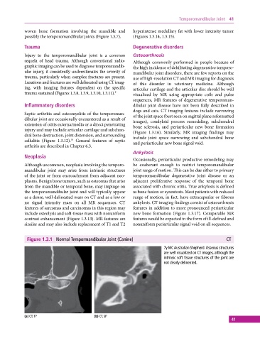
Figure 1.3.1 Normal Tempormandibular Joint (Canine) CT
7y MC Australian Shepherd. Osseous structures
are well visualized on CT images, although the
intrinsic soft tissue structures of the joint are
not clearly delineated.
(a) CT, TP (b) CT, SP
41