
Page 54 - Atlas of Small Animal CT and MRI
P. 54
44 Atlas of Small Animal CT and MRI
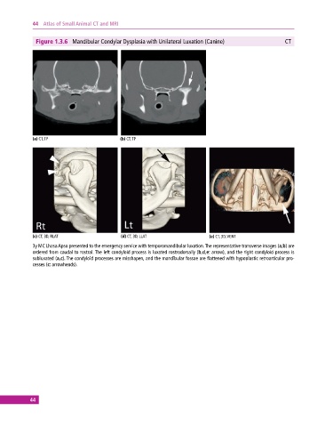
Figure 1.3.6 Mandibular Condylar Dysplasia with Unilateral Luxation (Canine) CT
(a) CT, TP (b) CT, TP
(c) CT, 3D, RLAT (d) CT, 3D, LLAT (e) CT, 3D, VENT
3y MC Lhasa Apso presented to the emergency service with temporomandibular luxation. The representative transverse images (a,b) are
ordered from caudal to rostral. The left condyloid process is luxated rostrodorsally (b,d,e: arrow), and the right condyloid process is
subluxated (a,c). The condyloid processes are misshapen, and the mandibular fossae are flattened with hypoplastic retroarticular pro-
cesses (c: arrowheads).
44