
Page 57 - Atlas of Small Animal CT and MRI
P. 57
Temporomandibular Joint 47
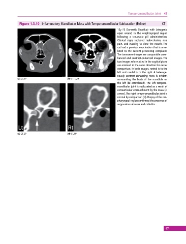
Figure 1.3.10 Inflammatory Mandibular Mass with Temporomandibular Subluxation (Feline) CT
12y FS Domestic Shorthair with iatrogenic
open wound in the oropharyngeal region
following a traumatic pill administration.
Clinical signs included malocclusion, oral
pain, and inability to close the mouth. The
cat had a previous enucleation that is unre-
lated to the current presenting complaint.
The transverse images are comparable unen-
hanced and contrast‐enhanced images. The
two images reformatted in the sagittal plane
are oriented in the same direction for easier
comparison. In both images, rostral is to the
left and caudal is to the right. A heteroge-
nously contrast‐enhancing mass is evident
(a) CT, TP (b) CT+C, TP surrounding the body of the mandible on
the left (b: arrowhead). The left temporo-
mandibular joint is subluxated as a result of
extraarticular encroachment by the mass (c:
arrow). The right temporomandibular joint is
normal by comparison (d). Biopsy of the oro-
pharyngeal region confirmed the presence of
suppurative abscess and cellulitis.
(c) CT, SP (d) CT, SP
47