
Page 62 - Atlas of Small Animal CT and MRI
P. 62
52 Atlas of Small Animal CT and MRI
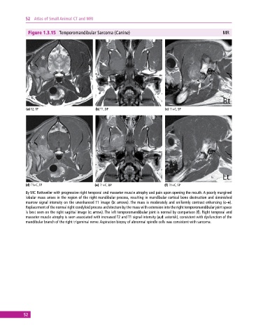
Figure 1.3.15 Temporomandibular Sarcoma (Canine) MR
(a) T2, TP (b) T1, DP (c) T1+C, SP
(d) T1+C, TP (e) T1+C, DP (f) T1+C, SP
8y MC Rottweiler with progressive right temporal and masseter muscle atrophy and pain upon opening the mouth. A poorly margined
lobular mass arises in the region of the right mandibular process, resulting in mandibular cortical bone destruction and diminished
marrow signal intensity on the unenhanced T1 image (b: arrows). The mass is moderately and uniformly contrast enhancing (c–e).
Replacement of the normal right condyloid process architecture by the mass with extension into the right temporomandibular joint space
is best seen on the right sagittal image (c: arrow). The left temporomandibular joint is normal by comparison (f). Right temporal and
masseter muscle atrophy is seen associated with increased T2 and T1 signal intensity (a,d: asterisk), consistent with dysfunction of the
mandibular branch of the right trigeminal nerve. Aspiration biopsy of abnormal spindle cells was consistent with sarcoma.
52