
Page 67 - Atlas of Small Animal CT and MRI
P. 67
Skull 57
imaging characteristics of these masses include T1 squamous cell carcinoma, can also involve the bones of
and T2 hypointensity with regions of hyperintensity. the skull (Figures 1.4.15, 1.4.16, 1.4.17). Lipomas or lipo-
Contrast enhancement is heterogeneous to uniform. 15 sarcomas have a characteristic fat attenuation (–100 HU)
Rarely, intracranial tumors, such as meningioma, within the musculature or soft tissues (Figure 1.4.18).
can expand outside the calvarium. Meningioma in Cats with pituitary adenomas may develop acromegaly
16
cats can also cause hyperostosis of the adjacent secondary to secretion of growth hormone and insulin‐
calvarium, and hyperostosis with bone lysis has been like growth factor. They tend to develop increased frontal
17
18
reported in the dog. Tumors of the soft tissues bone thickness and excess soft tissue in the nasal cavity,
surrounding the head, such as adenocarcinoma or sinuses, and pharynx, which can be seen on CT images. 1,19
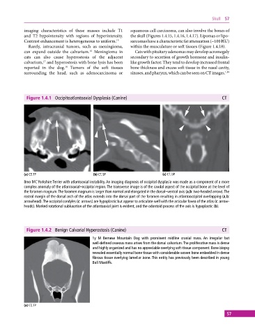
Figure 1.4.1 Occipitoatlantoaxial Dysplasia (Canine) CT
(a) CT, TP (b) CT, SP (c) CT, DP
8mo MC Yorkshire Terrier with atlantoaxial instability. An imaging diagnosis of occipital dysplasia was made as a component of a more
complex anomaly of the atlantoaxial–occipital region. The transverse image is of the caudal aspect of the occipital bone at the level of
the foramen magnum. The foramen magnum is larger than normal and elongated in the dorsal–ventral axis (a,b: two‐headed arrow). The
rostral margin of the dorsal arch of the atlas extends into the dorsal part of the foramen resulting in atlantooccipital overlapping (a,b:
arrowhead). The occipital condyles (c: arrows) are hypoplastic but appear to articulate well with the articular fovea of the atlas (c: arrow-
heads). Marked rotational subluxation of the atlantoaxial joint is evident, and the odontoid process of the axis is hypoplastic (b).
Figure 1.4.2 Benign Calvarial Hyperostosis (Canine) CT
1y M Bernese Mountain Dog with prominent midline cranial mass. An irregular but
well‐defined osseous mass arises from the dorsal calvarium. The proliferative mass is dense
and highly organized and has no appreciable overlying soft‐tissue component. Bone biopsy
revealed essentially normal bone tissue with considerable woven bone embedded in dense
fibrous tissue overlying lamellar bone. This entity has previously been described in young
Bull Mastiffs.
(a) CT, TP
57