
Page 69 - Atlas of Small Animal CT and MRI
P. 69
Skull 59
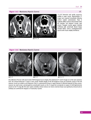
Figure 1.4.5 Masticatory Myositis (Canine) CT
1.5y MC Rottweiler with rapidly progressive
inability to open mouth. Representative CT
images were acquired immediately following
contrast medium administration. There is
moderate, diffuse contrast enhancement of the
left masseter and temporal muscles (a,b:
arrows). Pterygoid muscles appear relatively
unaffected (a,b: arrowhead). Muscle biopsy
revealed diffuse, chronic, lymphoplasmacytic
myositis with muscle atrophy and fibrosis.
(a) CT+C, TP (b) CT+C, TP
Figure 1.4.6 Masticatory Myositis (Canine) MR
(a) T2, TP (b) T1, TP (c) T1+C, TP
9mo Miniature Pinscher with recent onset of left temporal muscle atrophy. The unenhanced T1 and T2 images are at the same anatomic
level. The contrast‐enhanced T1 image is more caudal. Marked atrophy of the left temporal muscle and moderate atrophy of the left
masseter muscle are evident on all sequences. There is a pronounced increase in signal intensity of affected temporal (a: large arrows),
masseter (a: small arrow), and pterygoid (a: arrowheads) muscles on the T2 image that corresponds to regions of mild hyperintensity
present on the T1 image (b). The same regions markedly contrast enhance (c). Serum creatinine kinase was significantly elevated and an
antibody test confirmed the diagnosis of masticatory myositis.
59