Page 71 - Atlas of Small Animal CT and MRI
P. 71

Skull  61


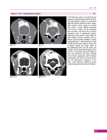
              Figure 1.4.9  Osteomyelitis (Canine)                                                        CT
                                                                             1y Pit Bull Terrier with an 8‐month history of
                                                                             progressive right‐sided head and facial swell-
                                                                             ing. Representative images are unenhanced
                                                                             (a,b) and contrast enhanced (c,d) at compa-
                                                                             rable anatomic levels. Images are ordered
                                                                             from  rostral to  caudal. Marked asymmetry
                                                                             of  the head is evident, and the underlying
                                                                             mass has both a soft tissue and an osseous
                                                                               component  (a,b).  A peripherally contrast‐
                                                                             enhancing cavitary lesion is seen within the
                                                                             right temporal muscle, consistent with an
                                                                             abscess and surrounding cellulitis (c). Dense
                                                                             osteoproliferation is seen involving the right
                                                                             and left parietal bones (a,b). An involucrum
            (a) CT, TP                       (b) CT, TP                      is  present toward the caudal aspect of
                                                                             the   proliferative bone mass (b: arrow) and
                                                                               contains a focal mineral‐dense body consist-
                                                                             ent with a sequestrum (d: arrow). Biopsy of
                                                                             bone and associated soft tissues revealed
                                                                             severe, chronic, suppurative, and necrotizing
                                                                             osteomyelitis with reactive new bone forma-
                                                                             tion.  The hyperostotic component of this
                                                                             lesion could represent underlying benign
                                                                               calvarial hyperostosis that became infected.


            (c) CT+C, TP                     (d) CT+C, TP


                                                                                                             61
   66   67   68   69   70   71   72   73   74   75   76