Page 72 - Atlas of Small Animal CT and MRI
P. 72

62  Atlas of Small Animal CT and MRI


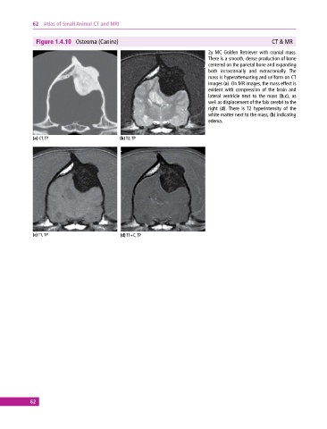
              Figure 1.4.10  Osteoma (Canine)                                                        CT & MR
                                                                              2y MC Golden Retriever with cranial mass.
                                                                              There is a smooth, dense production of bone
                                                                              centered on the parietal bone and expanding
                                                                              both intracranially and extracranially.  The
                                                                              mass is hyperattenuating and uniform on CT
                                                                              images (a). On MR images, the mass effect is
                                                                              evident  with  compression  of  the  brain  and
                                                                              lateral ventricle next to the mass (b,c), as
                                                                              well as displacement of the falx cerebri to the
                                                                              right (d).  There is  T2 hyperintensity of the
                                                                              white matter next to the mass, (b) indicating
                                                                              edema.


             (a) CT, TP                      (b) T2, TP


             (c) T1, TP                      (d) T1+C, TP


            62
   67   68   69   70   71   72   73   74   75   76   77