
Page 77 - Atlas of Small Animal CT and MRI
P. 77
Skull 67
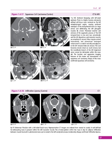
Figure 1.4.17 Squamous Cell Carcinoma (Canine) CT & MR
11y MC Shetland Sheepdog with left‐sided
epistaxis. There is a highly invasive and poorly
defined soft‐tissue mass centered on the left
retropharyngeal region, causing profound
osteolysis of the basisphenoid bone (a: large
arrow) and obliterating the left pterygoid
muscles (b: arrow). Periosteal reaction and
sclerosis of the zygomatic process of the left
temporal bone is also seen (a,c: arrowhead),
and the left digastricus and masseter muscles
are atrophied (c: arrows). Mass margins extend
intracranially, and pronounced meningeal
enhancement is evident ventrally and adjacent
to the left temporal lobe (d: arrows). The oval
(a) CT, TP (b) T2, TP foramina remain intact (a: small arrows), but
the mandibular branch of the left trigeminal
nerve cannot be delineated within the mass
(d). The location and aggressive imaging
features of this lesion are characteristic of
squamous cell carcinoma. Biopsy of the mass
confirmed squamous cell carcinoma.
(c) T1, TP (d) T1+C, TP
Figure 1.4.18 Infiltrative Lipoma (Canine) CT
(a) CT, TP (b) CT, TP (c) CT, TP
8y FS Doberman Pinscher with a left‐sided facial mass. Representative CT images are ordered from rostral to caudal. A well‐defined
fat‐attenuating mass is present within the left masseter muscle. The striated pattern within the mass is due to adipose infiltration
between muscle fasciculi A subchondral bone cyst is noted in the left condyloid process incidentally. Biopsy confirmed infiltrative lipoma.
67