
Page 81 - Atlas of Small Animal CT and MRI
P. 81
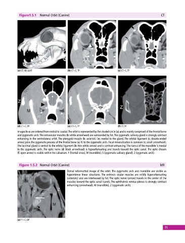
Figure1.5.1  Normal Orbit (Canine)                                                          CT


            (a) CT, 3D, LLAT                 (b) CT+C, TP                     (c) CT+C, TP


            (d) CT+C, TP                     (e) CT+C, TP                     (f) CT, TP
            Images b–e are ordered from rostral to caudal. The orbit is represented by the shaded circle (a) and is mainly comprised of the frontal bone
            and zygomatic arch. The extraocular muscles (b: white arrowhead) are surrounded by fat. The zygomatic salivary gland is strongly  contrast
            enhancing in the ventrolateral orbit. The pterygoid muscle (b: asterisk) lies medial to the gland. The orbital ligament (c: double‐ended
            arrow) joins the zygomatic process of the frontal bone (a: #) to the zygomatic arch. Focal mineralization is common (c: small arrowhead).
            The lacrimal gland is ventral to the orbital ligament (b: thin white arrow) and is contrast enhancing. The ramus of the mandible is medial
            to the zygomatic arch. The optic nerve (d: black arrowhead) is hypoattenuating and travels toward the optic canal. The optic chiasm
            (f: open arrow) is visible within the calvarium. F (frontal sinus), M (mandible), S (zygomatic salivary gland), Z (zygomatic arch).


              Figure 1.5.2  Normal Orbit (Canine)                                                        MR

                                             Dorsal reformatted image of the orbit. The zygomatic arch and mandible are visible as
                                             hypointense linear structures. The extrinsic ocular muscles are mildly hyperattenuating
                                             (asterisks) and are interleaved by fat. The optic nerve (arrow) travels in the center of the
                                             muscles toward the optic canal (caret). The ophthalmic venous plexus is strongly contrast
                                             enhancing (arrowhead). M (mandible), Z (zygomatic arch).


            (a) T1+C, DP

                                                                                                             71