
Page 85 - Atlas of Small Animal CT and MRI
P. 85
Orbit 75
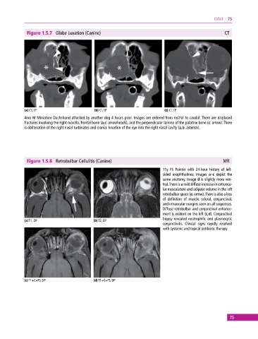
Figure 1.5.7 Globe Luxation (Canine) CT
(a) CT, TP (b) CT, TP (c) CT, TP
4mo M Miniature Dachshund attacked by another dog 4 hours prior. Images are ordered from rostral to caudal. There are displaced
fractures involving the right maxilla, frontal bone (a,c: arrowheads), and the perpendicular lamina of the palatine bone (c: arrow). There
is obliteration of the right nasal turbinates and cranial luxation of the eye into the right nasal cavity (a,b: asterisk).
Figure 1.5.8 Retrobulbar Cellulitis (Canine) MR
11y FS Pointer with 24‐hour history of left‐
sided exophthalmos. Images a–c depict the
same anatomy. Image d is slightly more ven-
tral. There is a mild diffuse increase in extraocu-
lar musculature and adipose volume in the left
retrobulbar space (a: arrow). There is also a loss
of definition of muscle, scleral, conjunctival,
and intraocular margins seen on all sequences.
Diffuse retrobulbar and conjunctival enhance-
ment is evident on the left (c,d). Conjunctival
biopsy revealed neutrophilic and plasmocytic
(a) T1, DP (b) T2, DP
conjunctivitis. Clinical signs rapidly resolved
with systemic and topical antibiotic therapy.
(c) T1+C+FS, DP (d) T1+C+FS, DP
75