
Page 89 - Atlas of Small Animal CT and MRI
P. 89
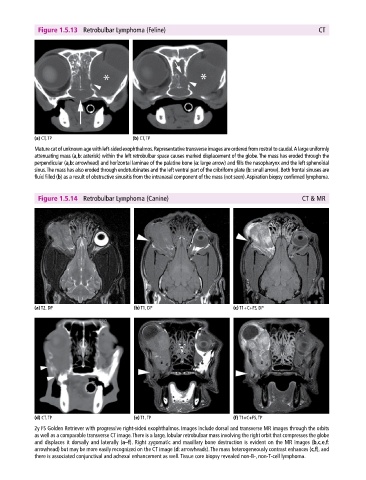
Figure 1.5.13  Retrobulbar Lymphoma (Feline)                                                CT


            (a) CT, TP                       (b) CT, TP
            Mature cat of unknown age with left‐sided exophthalmos. Representative transverse images are ordered from rostral to caudal. A large  uniformly
            attenuating mass (a,b: asterisk) within the left retrobulbar space causes marked displacement of the globe. The mass has eroded through the
            perpendicular (a,b: arrowhead) and horizontal laminae of the palatine bone (a: large arrow) and fills the nasopharynx and the left  sphenoidal
            sinus. The mass has also eroded through endoturbinates and the left ventral part of the cribriform plate (b: small arrow). Both frontal sinuses are
            fluid filled (b) as a result of obstructive sinusitis from the intranasal component of the mass (not seen). Aspiration biopsy confirmed lymphoma.


              Figure 1.5.14  Retrobulbar Lymphoma (Canine)                                           CT & MR


            (a) T2, DP                       (b) T1, DP                       (c) T1+C+FS, DP


            (d) CT, TP                       (e) T1, TP                       (f) T1+C+FS, TP

            2y FS Golden Retriever with progressive right‐sided exophthalmos. Images include dorsal and transverse MR images through the orbits
            as well as a comparable transverse CT image. There is a large, lobular retrobulbar mass involving the right orbit that compresses the globe
            and displaces it dorsally and laterally (a–f). Right zygomatic and maxillary bone destruction is evident on the MR images (b,c,e,f:
              arrowhead) but may be more easily recognized on the CT image (d: arrowheads). The mass heterogeneously contrast enhances (c,f), and
            there is associated conjunctival and adnexal enhancement as well. Tissue core biopsy revealed non‐B‐, non‐T‐cell lymphoma.