
Page 93 - Atlas of Small Animal CT and MRI
P. 93
Orbit 83
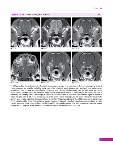
Figure 1.5.19 Orbital Meningioma (Canine) MR
(a) T1, TP (b) T2, TP (c) T1+C, TP
(d) T1+C, DP (e) T1+C, TP (f) T1+C, TP
7y MC Cavalier King Charles Spaniel with a 4‐month history of progressive right‐sided exophthalmos. The first three images (a–c) depict
the same anatomy and are at the level of the caudal aspect of the retrobulbar spaces. Images e and f are slightly more caudal and are
ordered from rostral to caudal with image f at the caudal‐most extent of the retrobulbar spaces. There is a well‐defined mass in the
caudal aspect of the right retrobulbar space that is hyperintense to gray matter on both T1 and T2 images (a,b: arrow). The mass is
moderately and uniformly contrast enhancing (c,d: arrowhead). The caudal extent of the mass is adjacent to the region of the orbital
fissure and optic canal (d–e,f: arrow). These structures are not clearly seen on the images, but the arrows indicate their approximate
location. Upon surgical exploration, the mass was found to arise from the ophthalmic branch of the right trigeminal nerve (cranial nerve
V) as it exited the orbital fissure. Excisional biopsy revealed meningioma. Although a specific preoperative diagnosis could not be made
from MR images, the caudal and central location of the mass within the retrobulbar space and the uniform contrast‐enhancement pattern
is suggestive of a neoplasm arising from one of the nerves arising from the orbital fissure or optic canal.
83