
Page 90 - Atlas of Small Animal CT and MRI
P. 90
80 Atlas of Small Animal CT and MRI
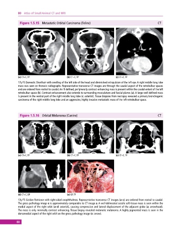
Figure 1.5.15 Metastatic Orbital Carcinoma (Feline) CT
(a) CT+C, TP (b) CT+C, TP (c) CT+C, TP
17y FS Domestic Shorthair with swelling of the left side of the head and diminished retropulsion of the left eye. A right middle lung lobe
mass was seen on thoracic radiographs. Representative transverse CT images are through the caudal aspect of the retrobulbar spaces
and are ordered from rostral to caudal. An ill‐defined, peripherally contrast‐enhancing mass is present within the caudal extent of the left
retrobulbar space (b). Contrast enhancement also extends to surrounding musculature and fascial planes (a). A large well‐defined mass
is present in the ventral part of the right middle lung lobe (c: asterisk). Tissue biopsies from necropsy revealed a primary bronchogenic
carcinoma of the right middle lung lobe and an aggressive, highly invasive metastatic mass of the left retrobulbar space.
Figure 1.5.16 Orbital Melanoma (Canine) CT
(a) CT+C, TP (b) CT+C, TP (c) CT+C, TP
(d) CT+C, DP (e) GP, TP
13y FS Golden Retriever with right‐sided exophthalmos. Representative transverse CT images (a–c) are ordered from rostral to caudal.
The gross pathology image e is approximately comparable to CT image a. A well‐delineated sessile soft‐tissue mass is seen within the
medial aspect of the right orbit (a–d: asterisk), causing compression and lateral displacement of the adjacent globe (a: arrowhead).
The mass is only minimally contrast enhancing. Tissue biopsy revealed melanotic melanoma. A highly pigmented mass is seen in the
dorsomedial aspect of the right orbit on the gross pathology image (e: arrow).
80