
Page 87 - Atlas of Small Animal CT and MRI
P. 87
Orbit 77
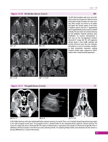
Figure 1.5.10 Retrobulbar Abscess (Canine) MR
10y MC Newfoundland with acute onset left‐
sided conjunctival hyperemia. Representative
dorsal plane images all depict the same anat-
omy. These images are ventral to the globes
and depict the ventral aspect of the orbits.
A fusiform abscess that has T1 hypointensity
and T2 hyperintensity (a–d: arrows) is evident
centrally. The core does not contrast enhance,
but the periphery intensely enhances (c,d).
Distribution of contrast enhancement is
best seen on the fat‐suppressed contrast‐
enhanced T1 image (d). The left zygomatic
salivary gland is also mildly enlarged and
contrast enhances more than the contralat-
(a) T1, DP (b) T2, DP eral gland as a result of secondary sialadeni-
tis (a,d: arrowhead). Aspiration cytology
revealed marked septic suppurative inflam-
mation with a mixed bacterial population.
(c) T1+C, DP (d) T1+C+FS, DP
Figure 1.5.11 Pterygoid Abscess (Canine) CT
(a) CT+C, TP (b) US, SP (c) CT+C, DP
3y M Golden Retriever with right‐sided buphthalmos and pain opening the mouth. There is an irregularly shaped, hypoattenuating region
in the right pterygoid muscle (a,c). The pterygoid muscle is located medial to the hyperattenuating zygomatic salivary gland on the
transverse images (a) and on ultrasound (b). The region of inflammation is hypoechoic and irregular on ultrasound images (b). There is
additional soft‐tissue opacity in the left nasal cavity indicating rhinitis. No migrating foreign bodies were observed, but this remains a
primary differential as a cause of the abscess.
77