
Page 82 - Atlas of Small Animal CT and MRI
P. 82
72 Atlas of Small Animal CT and MRI
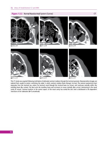
Figure 1.5.3 Normal Nasolacrimal System (Canine) CT
(a) CT+C, TP (b) CT+C, TP (c) CT+C, TP
(d) CT+C, TP (e) CT+C, TP (f) CT+C, TP
This CT study was acquired following instillation of iodinated contrast medium through the lacrimal punctum. Representative images are
ordered from caudal to rostral, mimicking the order in which contrast medium flows through the duct. The normal nasolacrimal duct
originates from the lacrimal sac, enters the lacrimal canal through the lacrimal bone (a: arrow), and continues rostrally within the
maxillary bone (b,c: arrow). The duct exits the maxillary bone and continues to course rostrally (d,e: arrow), terminating in the nasal
cavity (f: arrow). Contrast medium in the ventral aspect of the nasal cavity has exited the duct and is distributed in the dependent
interstices of the turbinates (b–f: arrowhead).
72