
Page 83 - Atlas of Small Animal CT and MRI
P. 83
Orbit 73
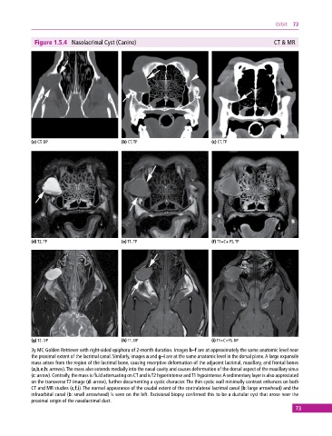
Figure 1.5.4 Nasolacrimal Cyst (Canine) CT & MR
(a) CT, DP (b) CT, TP (c) CT, TP
(d) T2, TP (e) T1, TP (f) T1+C+ FS, TP
(g) T2, DP (h) T1, DP (i) T1+C+FS, DP
3y MC Golden Retriever with right‐sided epiphora of 2‐month duration. Images b–f are at approximately the same anatomic level near
the proximal extent of the lacrimal canal. Similarly, images a and g–i are at the same anatomic level in the dorsal plane. A large expansile
mass arises from the region of the lacrimal bone, causing resorptive deformation of the adjacent lacrimal, maxillary, and frontal bones
(a,b,e,h: arrows). The mass also extends medially into the nasal cavity and causes deformation of the dorsal aspect of the maxillary sinus
(c: arrow). Centrally, the mass is fluid attenuating on CT and is T2 hyperintense and T1 hypointense. A sedimentary layer is also appreciated
on the transverse T2 image (d: arrow), further documenting a cystic character. The thin cystic wall minimally contrast enhances on both
CT and MR studies (c,f,i). The normal appearance of the caudal extent of the contralateral lacrimal canal (b: large arrowhead) and the
infraorbital canal (b: small arrowhead) is seen on the left. Excisional biopsy confirmed this to be a ductular cyst that arose near the
proximal origin of the nasolacrimal duct.
73