
Page 88 - Atlas of Small Animal CT and MRI
P. 88
78 Atlas of Small Animal CT and MRI
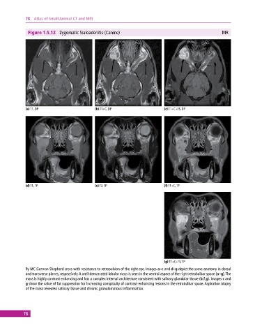
Figure 1.5.12 Zygomatic Sialoadenitis (Canine) MR
(a) T1, DP (b) T1+C, DP (c) T1+C+FS, DP
(d) T1, TP (e) T2, TP (f) T1+C, TP
(g) T1+C+FS, TP
8y MC German Shepherd cross with resistance to retropulsion of the right eye. Images a–c and d–g depict the same anatomy in dorsal
and transverse planes, respectively. A well‐demarcated lobular mass is seen in the ventral aspect of the right retrobulbar space (a–g). The
mass is highly contrast enhancing and has a complex internal architecture consistent with salivary glandular tissue (b,f,g). Images c and
g show the value of fat suppression for increasing conspicuity of contrast‐enhancing lesions in the retrobulbar space. Aspiration biopsy
of the mass revealed salivary tissue and chronic granulomatous inflammation.
78