
Page 92 - Atlas of Small Animal CT and MRI
P. 92
82 Atlas of Small Animal CT and MRI
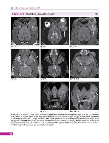
Figure 1.5.18 Orbital Rhabdomyosarcoma (Canine) MR
(a) T1, DP (b) T2, DP (c) T1+C+FS, DP
(d) T1, DP (e) T2, DP (f) T1+C+FS, DP
(g) T2, TP (h) T1+C+FS, TP
3y MC Tibetan Terrier with a 2‐week history of left‐sided exophthalmos. Representative dorsal plane images are at (a–c) and ventral to
(d–f) the level of the eyes. There is a large irregularly shaped mass within the retrobulbar space (a–c,g,h: asterisk), which is isointense
to gray matter on both T1 and T2 images. The mass invades the left temporal muscle (a–c: arrow) and appears to cause osteolysis of the
left zygomatic bone (d,h: arrowheads). The mass is nonuniformly contrast enhancing. Incidentally, the optic chiasm and origins of the
optic nerves are particularly well seen in this study (e,f: arrow), and the origin of the left optic nerve does not appear to be involved.
Histologic diagnosis following necropsy was rhabdomyosarcoma.
82