
Page 84 - Atlas of Small Animal CT and MRI
P. 84
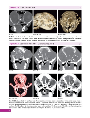
Figure 1.5.5  Orbital Fracture (Feline)                                                      CT


             (a) CT, TP                       (b) CT, 3D, RLAT                 (c) CT, 3D, CRAN
             2y MC Domestic Shorthair with acute head trauma of unknown cause. There is a moderately displaced fracture of the right ventromedial
             orbit (a–c: arrow). The fracture line is in the region of the convergence of the maxillary, lacrimal, and zygomatic bones. The cat also
               sustained a displaced fracture of the hard palate and separations of the incisive and mandibular symphyses (c: arrowheads).

              Figure 1.5.6  Deformation of the Orbit—Chronic Trauma (Canine)                               CT


             (a) CT, TP                       (b) CT, TP                       (c) CT, TP


             (d) CT, 3D, CRAN                 (e) CT+C, TP                     (f) CT+C, TP
             16mo M Chihuahua bitten in the face 1 year prior. The representative transverse images a–c are ordered from rostral to caudal. Images e
             and f are contrast‐enhanced images comparable to b and c, respectively. There is marked deformation of the right maxilla and frontal
             bone (a,b: arrowheads) and multiple fenestrations within the right maxilla and left frontal bone (b,c: arrows), causing both orbits to be
             misshapen. The 3D rendering (d) shows the extent of trauma and remodeling of the dorsal margin of the right orbit. Right‐sided phthisis
             bulbi (e: arrow) and an intracranial cyst (f: asterisk) resulting from the previous trauma are also evident.
            74