
Page 98 - Atlas of Small Animal CT and MRI
P. 98
88 Atlas of Small Animal CT and MRI
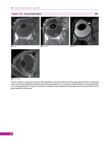
Figure 1.6.2 Normal Globe (Feline) MR
(a) T1, DP (b) T1+C, DP (c) T2, DP
(d) T1+C+FS, SP
The lens is visible as a hypointense structure with a hyperintense capsule (b: asterisk). The ciliary body supports the lens (c: arrowhead)
and is contrast enhancing (d). The anterior (b: small white arrow), posterior (d: <), and vitreous chambers (b: open arrow) are hypointense
on T1 and hyperintense on T2 images. The cornea (d: arrowhead) is visible anteriorly. The enhancing retina (b: large solid white arrow) is
present posterior to the vitreous.
88