
Page 101 - Atlas of Small Animal CT and MRI
P. 101
Globe 91
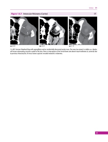
Figure 1.6.7 Intraocular Melanoma (Canine) CT
(a) CT, TP (b) CT, TP (c) CT, TP
11y MC German Shepherd Dog with aspergillosis and an incidentally discovered ocular mass. The mass (a: arrows) is visible as a lobular,
soft‐tissue attenuating structure caudal to the lens. There is interruption of the frontal bone and absent nasal turbinates (c: asterisk) due
to previous rhinosinusitis. Vitreous humor aspirate revealed melanotic melanoma.
91